


Home / Training / Manuals / Atlas of breast cancer early detection / Cases
Atlas of breast cancer early detection
Go back to the list of case studies
.png) Click on the pictures to magnify and display the legends
Click on the pictures to magnify and display the legends
| Case number: | 038 |
| Age: | 74 |
| Clinical presentation: | Postmenopausal woman with increased risk of developing breast cancer presented for screening. Examination did not reveal any significant lump or irregularity in the breasts or axillae. |
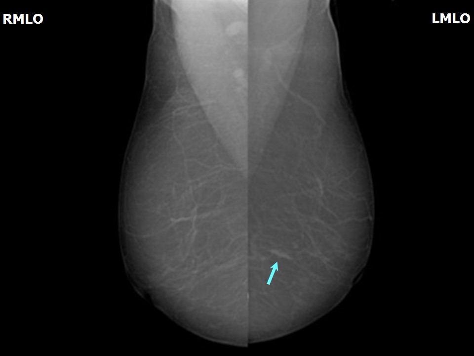
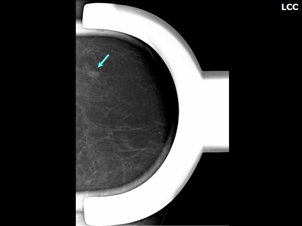
Mammography:
 |  |
 |
| Breast composition: | ACR category a (the breasts are almost entirely fatty) | Mammography features: |
| ‣ Location of the lesion: | Left breast, lower outer quadrant at 4 oclock, middle and posterior thirds |
| ‣ Mass: | |
| • Number: | 1 |
| • Size: | 0.6 cm in greatest dimension |
| • Shape: | Irregular |
| • Margins: | Indistinct |
| • Density: | High |
| ‣ Calcifications: | |
| • Typically benign: | None |
| • Suspicious: | None |
| • Distribution: | None |
| ‣ Architectural distortion: | None |
| ‣ Asymmetry: | Focal |
| ‣ Intramammary node: | None |
| ‣ Skin lesion: | None |
| ‣ Solitary dilated duct: | None |
| ‣ Associated features: | None |
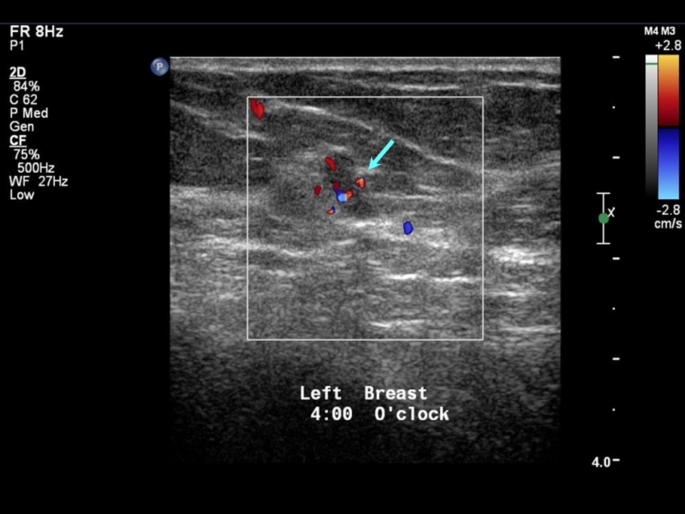
Ultrasound:
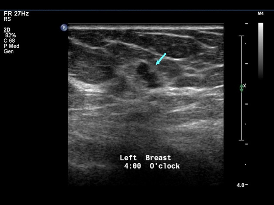 |  |
| Ultrasound features: Left breast, lower outer quadrant at 4 oclock | |
| ‣ Mass | |
| • Location: | Left breast, lower outer quadrant at 4 oclock |
| • Number: | 1 |
| • Size: | 0.6 cm in greatest dimension |
| • Shape: | Irregular |
| • Orientation: | Not parallel |
| • Margins: | Angular |
| • Echo pattern: | Hypoechoic |
| • Posterior features: | No posterior features |
| ‣ Calcifications: | None |
| ‣ Associated features: | Internal vascularity |
| ‣ Special cases: | None |
BI-RADS:
BI-RADS Category: 4B (moderate suspicion of malignancy)Further assessment:
Further assessment advised: Referral for excision biopsyCytology:
 |
| Cytology features: | |
| ‣ Type of sample: | FNAC |
| ‣ Site of biopsy: | |
| • Laterality: | |
| • Quadrant: | |
| • Localization technique: | Palpation |
| • Nature of aspirate: | |
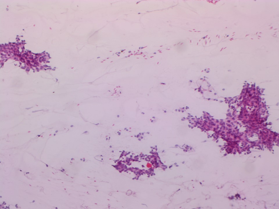
| ‣ Cytological description: | Smears show many fibroadipose tissue fragments and a few cohesive clusters of benign ductal epithelial cells against a haemorrhagic background |
| ‣ Reporting category: | Benign |
| ‣ Diagnosis: | Benign proliferative breast lesion |
| ‣ Comments: | None |
Histopathology:
Lumpectomy
 |
| Histopathology features: | |
| ‣ Specimen type: | Lumpectomy |
| ‣ Laterality: | |
| ‣ Macroscopy: | Specimen (7.0 × 8.0 × 4.0 cm) with skin flap (6.0 cm in length). A small whitish area (0.4 cm in diameter) is seen 2.0 cm from the superior margin. The rest of the breast parenchyma is unremarkable and no other suspicious nodular areas are seen on gross examination |
| ‣ Histological type: | Section from the whitish area shows multiple dilated cystic ducts lined by metaplastic apocrine epithelium with lumen containing thick proteinaceous material. There are also ducts showing epithelial hyperplasia. One of the ducts shows papillary hyperplasia with a few micropapillae. There is no evidence of nuclear atypia and DCIS. Sections were taken from all firm-appearing areas in the lumpectomy specimen. Section 1189H/14 shows two ducts with atypical cribriform hyperplasia. All remaining sections show foci with mild to moderate epithelial hyperplasia and columnar cell hyperplasia |
| ‣ Histological grade: | |
| ‣ Mitosis: | |
| ‣ Maximum invasive tumour size: | |
| ‣ Lymph node status: | |
| ‣ Peritumoural lymphovascular invasion: | |
| ‣ DCIS/EIC: | Absent |
| ‣ Margins: | Section from the lateral margin of the lumpectomy shows a few ducts with moderate epithelial hyperplasia. Sections from superior, medial, and inferior margins and base show only fibroadipose tissue |
| ‣ Pathological stage: | |
| ‣ Biomarkers: | |
| ‣ Comments: |
Case summary:
| Postmenopausal woman with increased risk of developing breast cancer presented for mammography screening. Diagnosed as focal asymmetry in left breast, BI-RADS 4B on imaging, as benign proliferative breast lesion on cytology, and as duct with papillary hyperplasia with a few micropapillae and atypical cribriform hyperplasia on histopathology. |
Learning points:
|